3d/4d Ultrasound
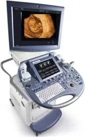
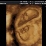
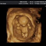
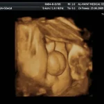
3D and 4D ultrasound rearrange and synthesize images taken of any part of the body that has 3D or 4D dimensions to create a similar size within the device. Like regular ultrasound(2D), 3D and 4D ultrasound are used to create a picture of your child in the womb, the difference between them is that 3D ultrasound creates a 3D image of your child, by taking multiple 2D images at different angles and then grouped together to form a 3D view. While 4D ultrasound creates a direct video effect, like a movie - you can watch your baby smile or yawn. Al-Hayat Medical Center in Qatar is distinguished by the availability of the best 3D and 4D ultrasound devices, as they are currently considered an essential way for doctors to verify the health of the fetus and discover the presence of any congenital malformations, disabilities or chromosomal abnormalities (chromosome), whether it is related to the surface, viscera, or structure of the fetus. Not forgetting to mention, the real pleasure that parents gain in examining the images of stereoscopic embryos, as they move in front of them in "real-time" when using three-dimensional and four-dimensional ultrasound. This is of great benefit in strengthening the relationship between parents and children as well as accepting birth defects (if any) and dealing with is more rationally and alleviating the symptoms of psychological trauma that follow the birth. The pregnant woman can obtain a video or DVD documenting her examination using the full 3D and 4D ultrasound, in addition to pictures of her fetus to start his first album.
Indications for 3D/4D ultrasound:
3D/4D ultrasound has many indications including:
1- Follow up of pregnancy and fetal wellbeing in different stages of pregnancy:
A - First trimester:
-
Determination of gestational age.
-
Seeing fetal heart activity.
-
Determination of the number of fetuses.
-
Determination of pregnancy location either intrauterine or extrauterine.
-
Cases of vaginal bleeding (threatened abortion).
-
Detection of neural tube defects (skull and spine).
-
Diagnosis of cases of Mongolian children (Down syndrome).
-
See the upper and lower extremities and facial configurations
-
Determination of fetal sex.
B - 2nd and 3rd trimesters:
-
Detailed cranium and brain examination.
-
Detection of internal organs anomalies: heart, brain, lungs, kidneys, stomach, esophagus, bladder, and liver.
-
Examination of the chest, thoracic cage, and abdomen.
-
Identification of any chromosomal (chromosomal) or congenital malformations
-
Assessment of fetal growth, development, and movements, and coordination.
-
Determination of the placenta position and any associated problems such as limitation and infarction.
-
Assessment of amniotic fluid and blood flow velocity in the umbilical arteries using Doppler.
-
Assessment of fetal look, behavior, and environment.
-
Assessment of the risk of preterm delivery.
2-The use of 3D/4D ultrasound in gynecology and infertility:
-
Precise determination of ovarian size.
-
Measurement of the number and size of the follicles (cycle monitoring).
-
Diagnosis of polycystic ovaries.
-
Follow up of follicular size and detection of ovarian hyperstimulation in cases of ovulation induction using gonadotropins.
-
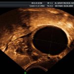
Diagnosis of ovarian cysts, tumors, and cancers.
-
Distinguish fluid accumulation in fallopian tubes and ovarian cysts
-
Assessment of tubal patency using a normal saline injection in the uterine cavity.
-
Measurement of endometrial thickness - Diagnosis of uterine anomalies - Detection of other uterine problems such as febrile fibers and tumorsز
-
Determining the location of the IUD in the womb, especially the hormonal IUD, which is difficult to view using regular devices.
-
Diagnosis of tumors and distinguishing malignant from benign, especially in the breasts, testes, uterus, ovary, and cervix. This is supported by the use of Doppler, which shows the degree of blood nutrition that reaches this tumor.
-
Periodic breast examination and distinguishing any tubers or clumps
-
Managing infertility in men by examining the testicles and checking for the presence of varicocele or fluid accumulation in the scrotum (hydrocele) and examination of the prostate gland.
Frequently asked questions on 3D/4D ultrasound device:
1- What do you mean by 3D? And what is 4D?
When we talk about 3D (3 dimensions) we mean the length, with and height. The fourth dimension is the time, which gives life to the objects. In 3D ultrasound machines, the operator has to spend the effort to get the 3rd dimension which requires a little more time; while in the 4D machines the 3rd dimension is automatically obtained by the machine itself in different but successive times giving life to the image.
2- What are the precautions before performing 3D/4D ultrasonography?
All you need is an appointment, but sometimes your bladder should be full, especially in women who prefer abdominal rather than vaginal examination. In this situation, you should drink 4 cups of water or juice and wait for a while before the examination.
3- Is it a safe examination? And how much time does it require?
Yes, it is, it is very safe and it requires 10-20 minutes.
4- When we consider the 3D/4D ultrasound examination very important?
This examination is recommended for all pregnant women but some of them need it more than others (advanced age, the previous baby with congenital anomalies, negative blood tests, family history of congenital anomalies, maternal exposure to teratogenic drugs or inhalation of toxic materials…. etc.).
5- What is the best time for 3D/4D ultrasound during pregnancy?
3D/4D ultrasound can be performed anytime and every visit during pregnancy. The minimum requirement is three examinations:
-
At 12-14 weeks: for detection of some malformations such as anencephaly, spina bifida, and malformations of the abdominal wall and extremities.
-
At 20-24 weeks: for searching in more details of the fetus and studying the internal organs and their functions, the most important of which is the heart.
-
At 30-34 weeks: for evaluation of fetal growth, fetal movements, and muscular coordination.
6- Does the 3D and 4D ultrasound scan substitute amniotic amniocentesis?
No, amniocentesis is useful in identifying problems in the structure of fetal cells (such as chromosomal abnormalities), while examination with a three-dimensional and four-dimensional ultrasound device reveals the largest problems at the level of the fetal organs themselves.
7- When is the best time to get the best images when scanning with 3D and 4D ultrasound?
It is possible to get great pictures of the fetus at all stages of pregnancy, and in general, the most beautiful pictures can be taken for the fetus between the 20 and 30 weeks of pregnancy.
8- Is there any guarantee to get good images as we can see on your website?
The image quality depends on many factors including the thickness of the abdominal wall of the pregnant woman, position of the fetus in the uterine cavity, gestational age, and amount of amniotic fluid around the fetus and, finally, experience of the sonographer. We can give a guarantee for the last factor only (experience).
8- Can I get the photos and DVD?
Yes, of course, just before leaving the clinic